
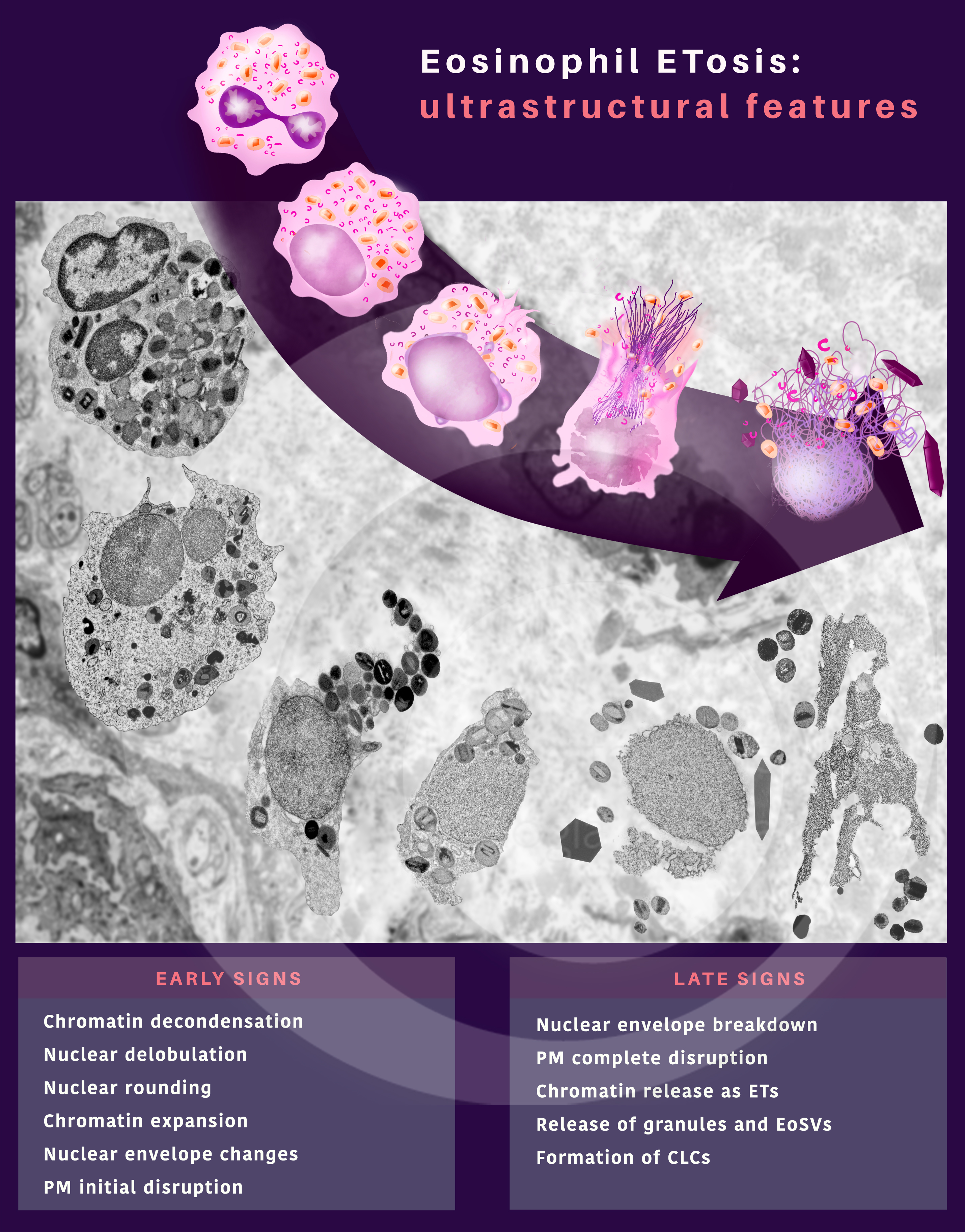
 Alterações ultraestruturais durante o processo ETosis de eosinófilos humanos in vivo
–
NEVES, V. H. et al. (2022) In Vivo ETosis of Human Eosinophils: The Ultrastructural Signature Captured by TEM in Eosinophilic Diseases. Front Immunol.
–
 Eventos da mitofagia em eosinófilos imaturos
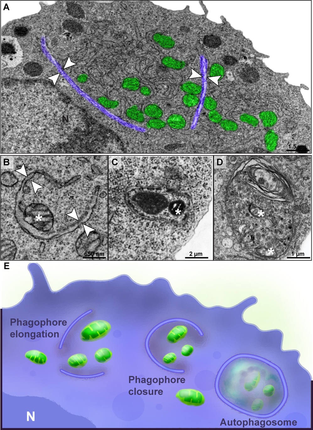
BONJOUR, K. et al. (2022) Mitochondrial Population in Mouse Eosinophils: Ultrastructural Dynamics in Cell Differentiation and Inflammatory Diseases. Front Cell Dev Biol.
–
 Estágios do desenvolvimento de eosinófilos observados pela Microscopia Eletrônica de Transmissão
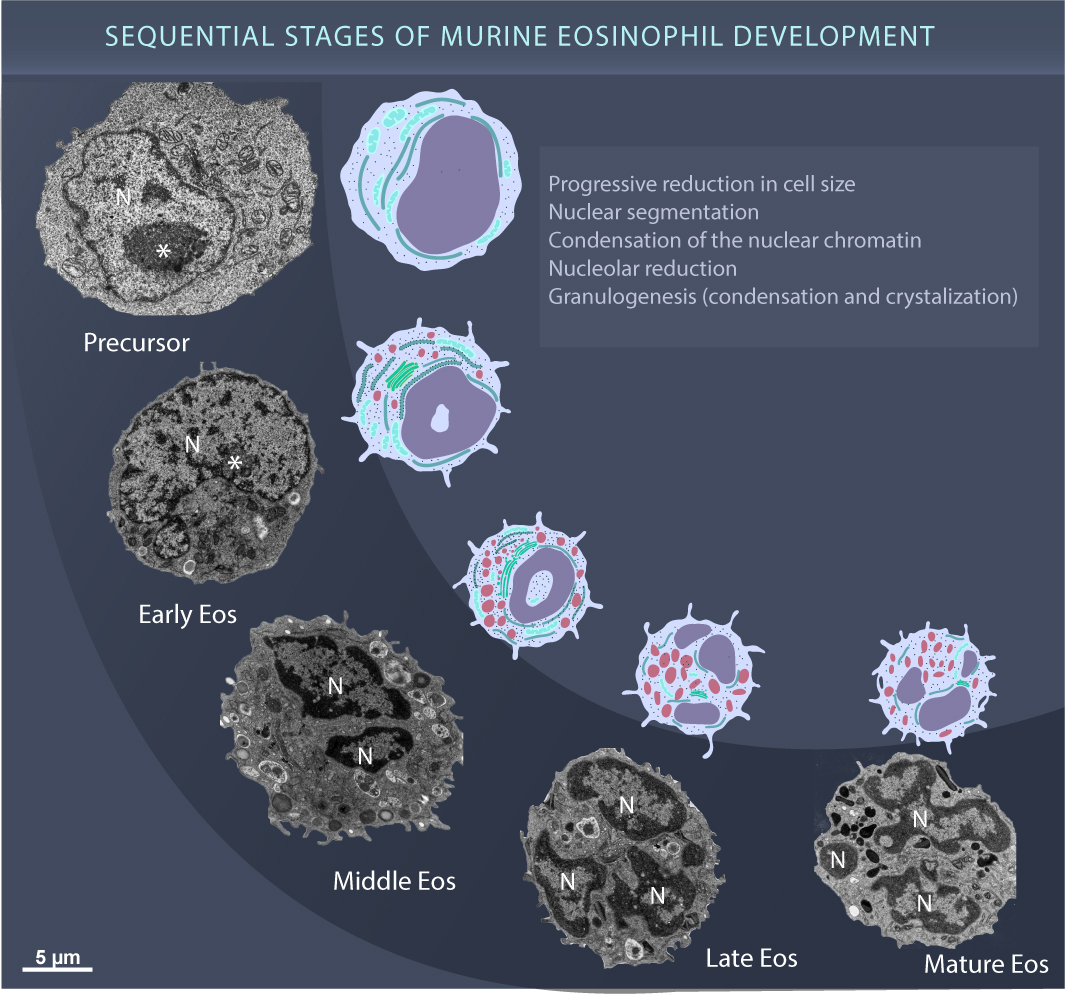
BONJOUR, K. et al. (2022) Mitochondrial Population in Mouse Eosinophils: Ultrastructural Dynamics in Cell Differentiation and Inflammatory Diseases. Front Cell Dev Biol.
–
 Sucessão ecológica no granuloma hepático de Schistosoma mansoni
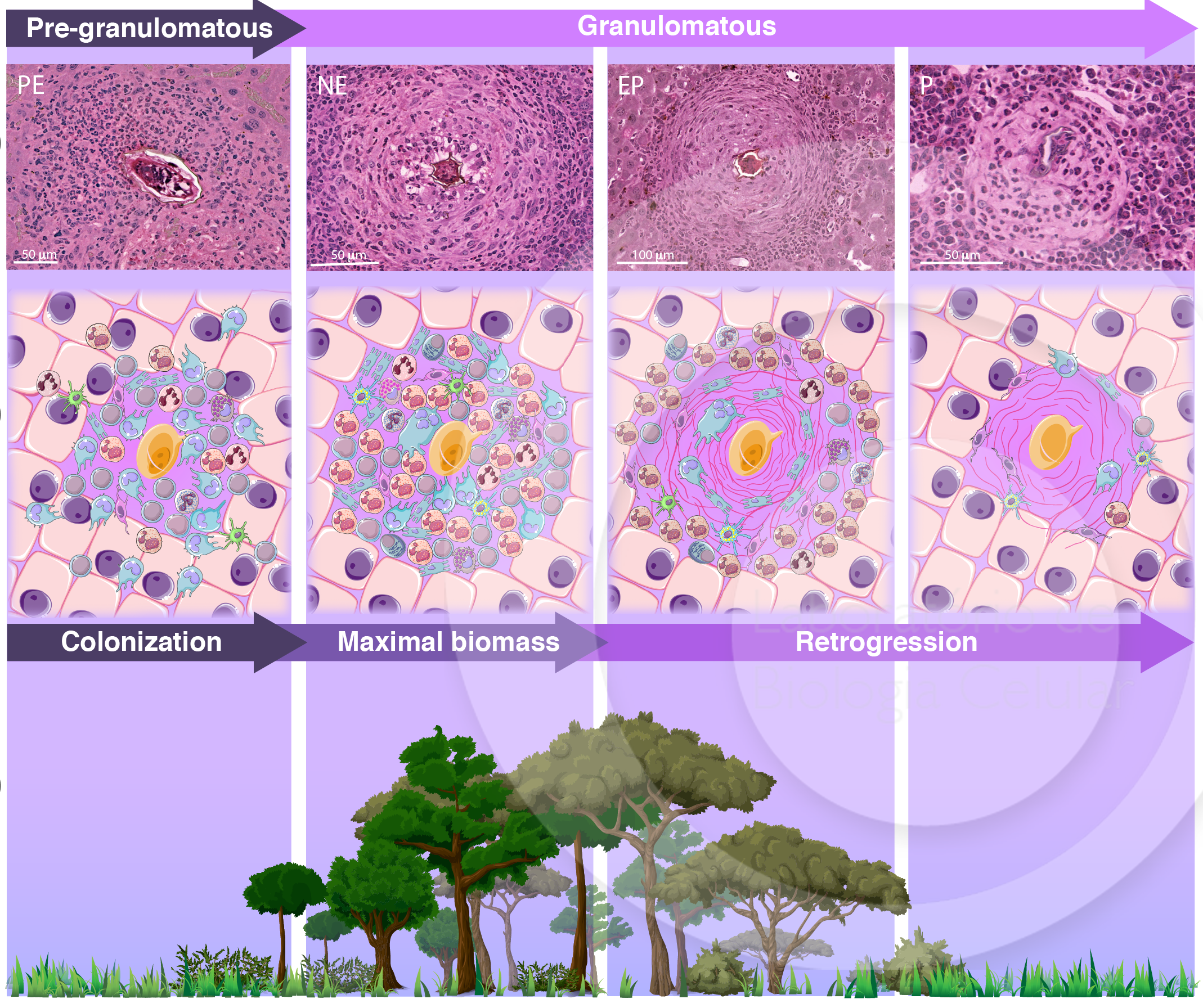
MALTA, K. K. et al. (2021) Changing our view of the Schistosoma granuloma to an ecological standpoint. Biol Rev.
–
 Whole slide imaging de granulomas em uma secção de fígado infectado com Schistosoma mansoni
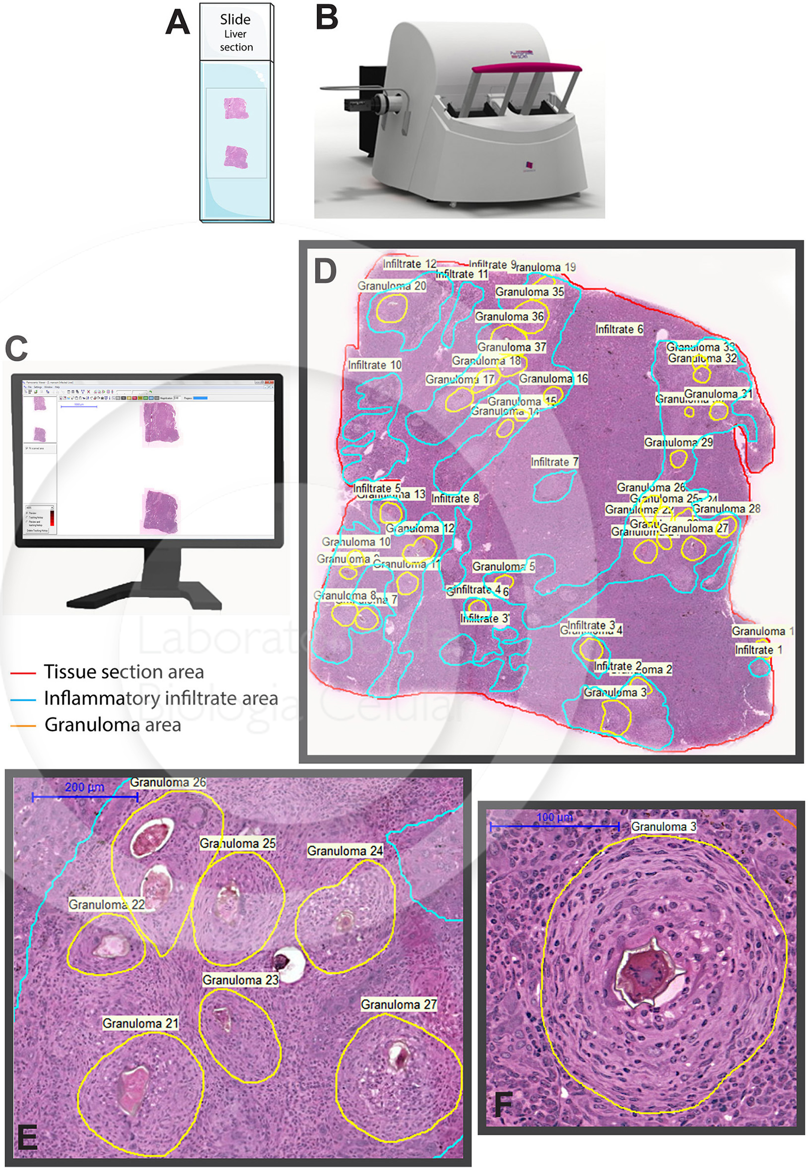
MELO, R. C. N. et al. (2020) Whole slide imaging and its applications to histopathological studies of liver disorders. Front Med.
–
 Morfologia das vesículas secretoras de eosinófilos humanos (Eosinophil Sombrero Vesicles)
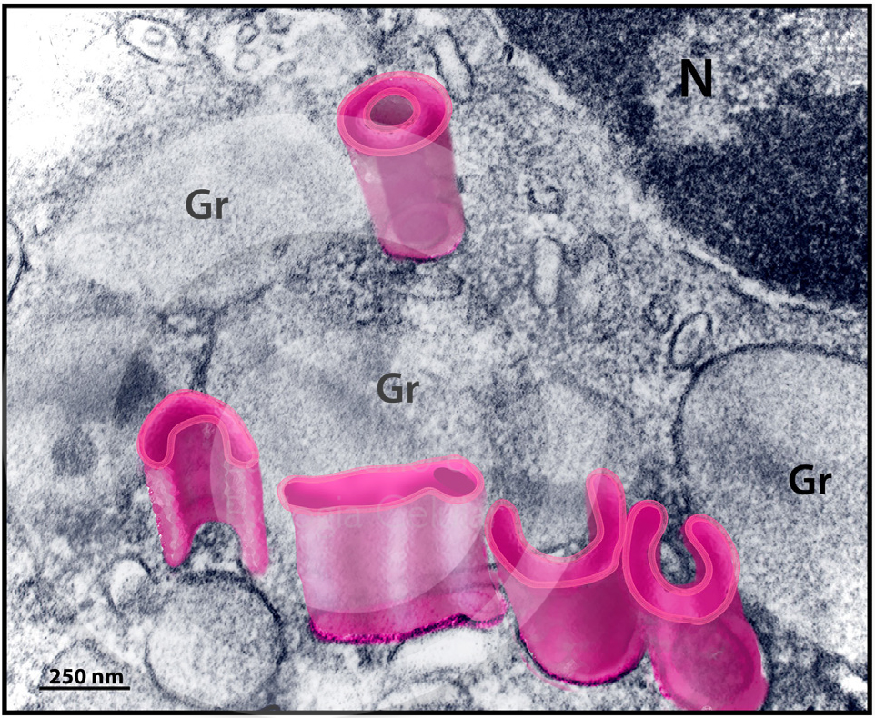
CARMO, L. A. S. et al. (2018) Single-Cell Analyses of Human Eosinophils at High Resolution to Understand Compartmentalization and Vesicular Trafficking of Interferon-Gamma. Front Immunol.
–
 Estágios de desenvolvimento do grânulo secretor de eosinófilos humanos
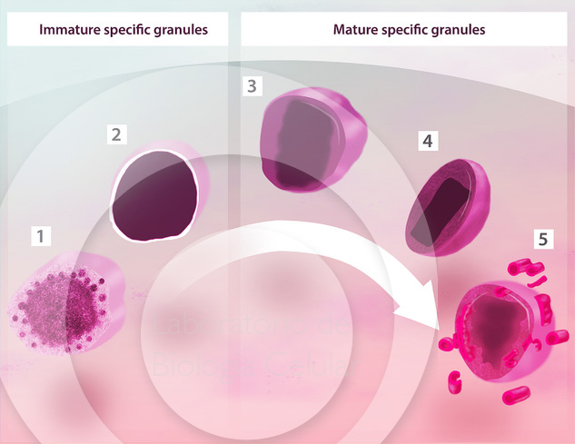
MELO, R. C. N.; WELLER, P. F. (2018). Contemporary understanding of the secretory granules in human eosinophils. J Leukoc Biol.
–
 Liberação de vesículas de membrana externa por bactérias de ecossistemas aquáticos
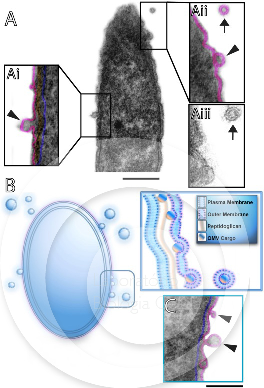
GAMALIER, J. P. et al. (2017). Increased production of outer membrane vesicles by cultured freshwater bacteria in response to ultraviolet radiation. Microbiol Res.
–
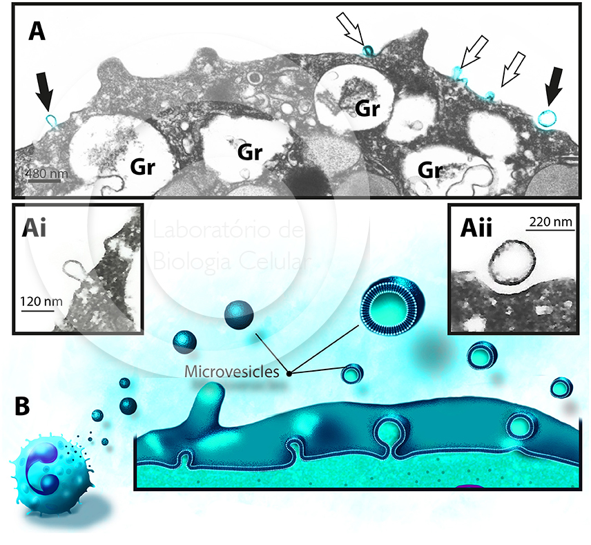
Liberação de micro vesículas por eosinófilo humano ativado por estímulos inflamatórios
AKUTHOTA, P. et al. (2016). Extracellular Microvesicle Production by Human Eosinophils Activated by “Inflammatory” Stimuli. Front Cell Dev Biol.
–
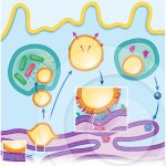 Um modelo que explica o papel dos corpúsculos lipídicos durante infecções com diferentes patógenos intracelulares
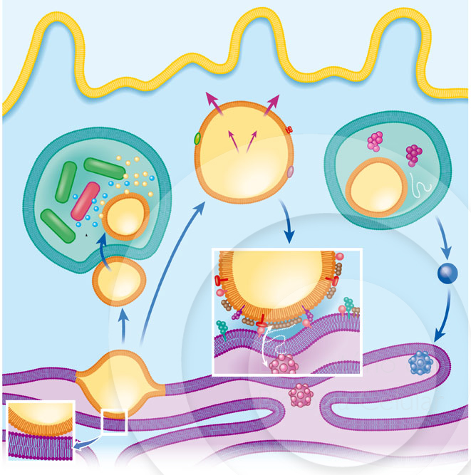
ROINGEARD, P.; MELO, R. C. N. (2016). Lipid droplet hijacking by intracellular pathogens. Cell Microbiol.
–
 Corpúsculos lipídicos se acumulam no citoplasma da célula hospedeira em resposta à interação com parasitas protozoários e favorecem a sobrevivência do parasito
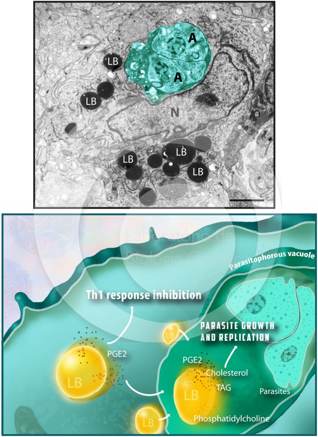
TOLEDO, D. A. et al. (2016). Host Lipid Bodies as Platforms for Intracellular Survival of Protozoan Parasites. Front Immunol.
–
 Formação de corpúsculos lipídicos induzida por diversos patógenos
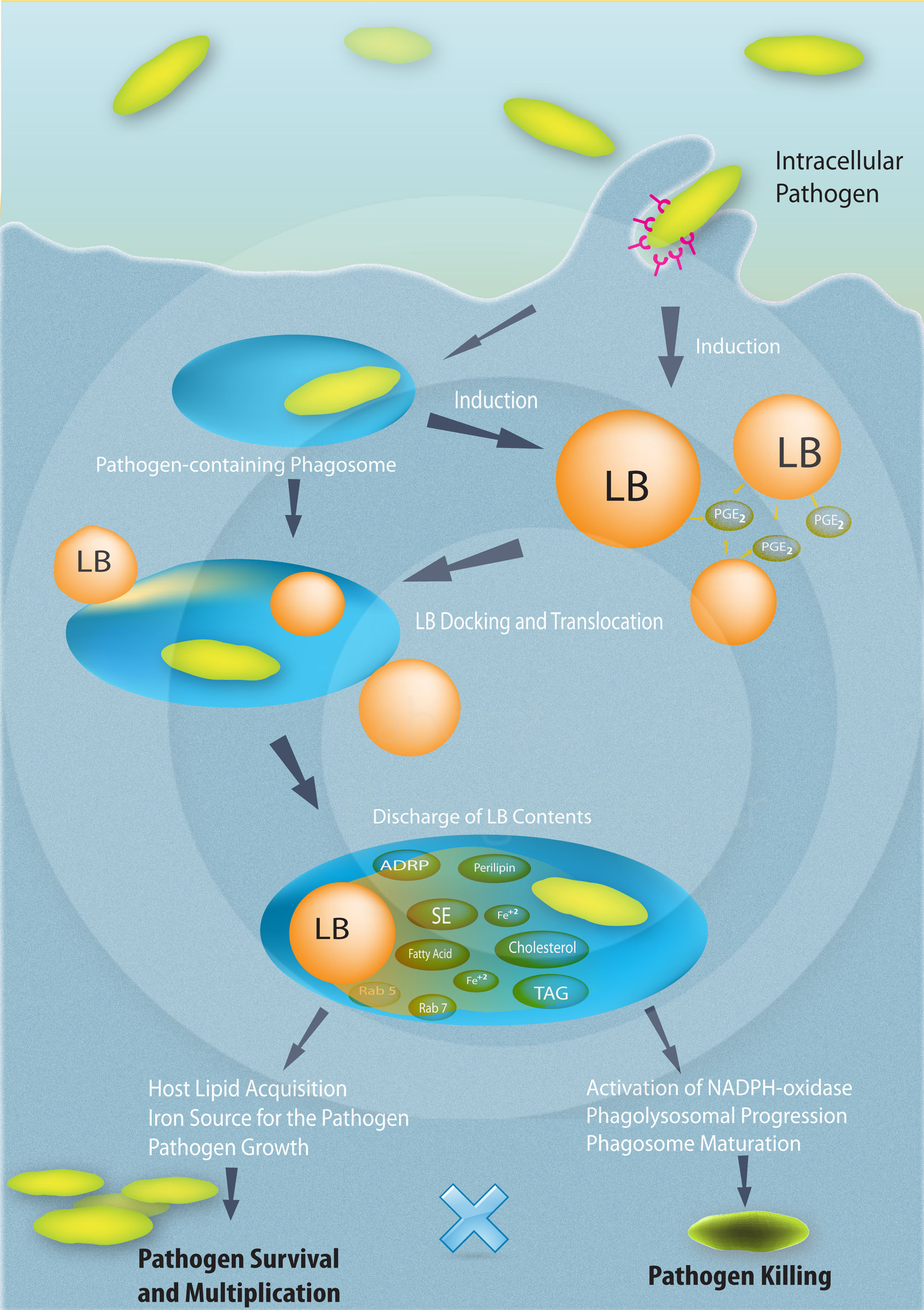
DIAS, F. F. et al. (2014). The Intriguing Ultrastructure of Lipid Body Organelles Within Activated Macrophages. Microsc Microanal.
–
 Aumento da expressão de PPARγ e acúmulo de lipídeos durante a infecção por M. bovis BCG
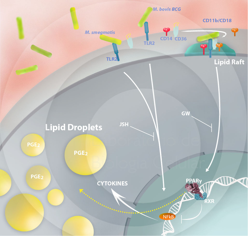
ALMEIDA, P. E. et. al. (2014). Differential TLR2 downstream signaling regulates lipid metabolism and 2 cytokine production triggered by Mycobacterium bovis BCG infection. Biochimica et Biophysica Acta.
–
 Efeitos biológicos das citocinas derivadas do eosinófilo na saúde e na doença
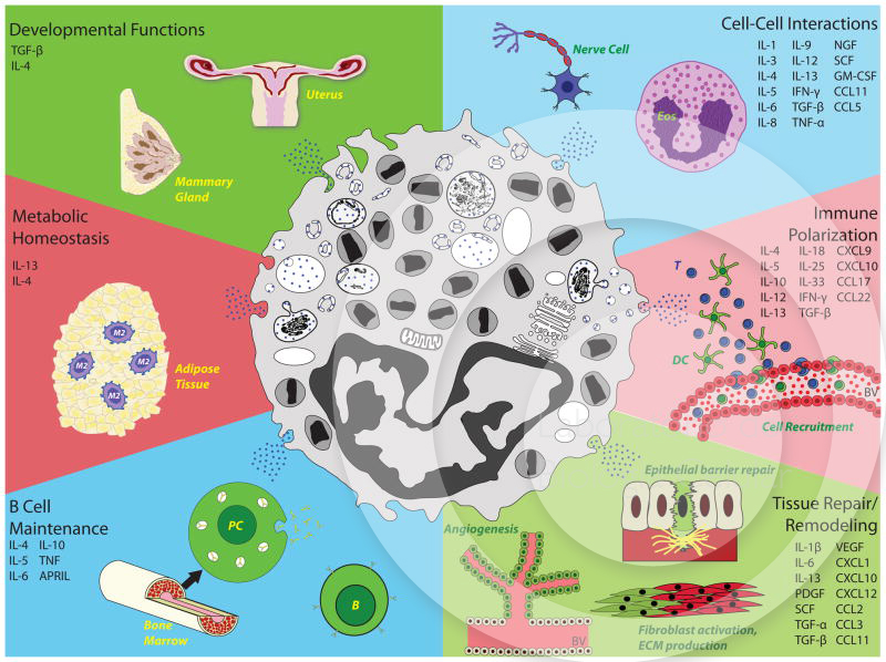
MELO, R. C. N. et al. (2013). Eosinophil-derived cytokines in health and disease: unraveling novel mechanisms of selective secretion. Allergy.
–
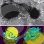 Arquitetura tridimensional (3D) de corpúsculos lipídicos em eosinófilos humanos
MELO, R. C. N. et al. (2013). The internal architecture of leukocyte lipid body organelles captured by three-dimensional electron microscopy tomography. Plos One.
–
 Corpúsculo lipídico, estrutura e composição
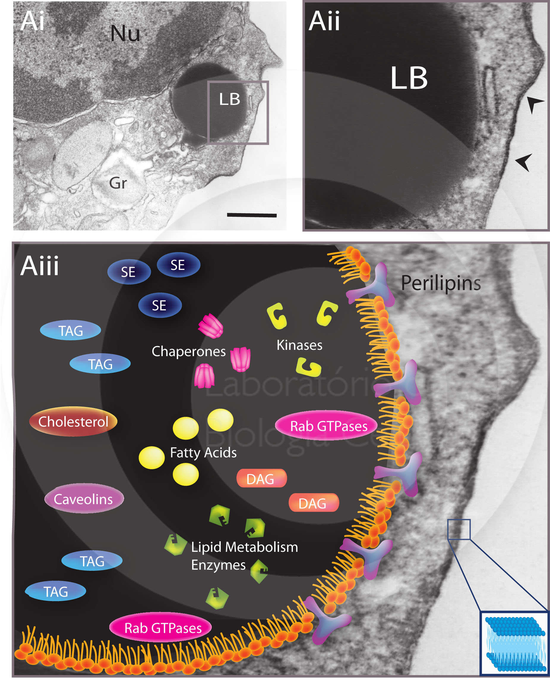
MELO, R. C. N; DVORAK, A. M. (2012). Lipid Body–Phagosome Interaction in Macrophages during Infectious Diseases: Host Defense or Pathogen Survival Strategy? PLoS Pathog.
–
 Comparação entre a membrana do corpúsculo lipídico e da membrana plasmática
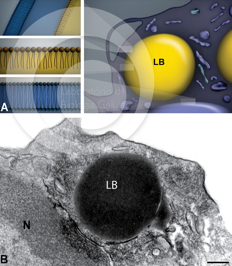
MELO, R. C. N. et al. (2011). Lipid bodies in inflammatory cells: structure, function, and current imaging techniques. J Histochem Cytochem.
–
 Ativação de macrófagos inflamatórios do coração durante a infecção aguda pelo Trypanosoma cruzi
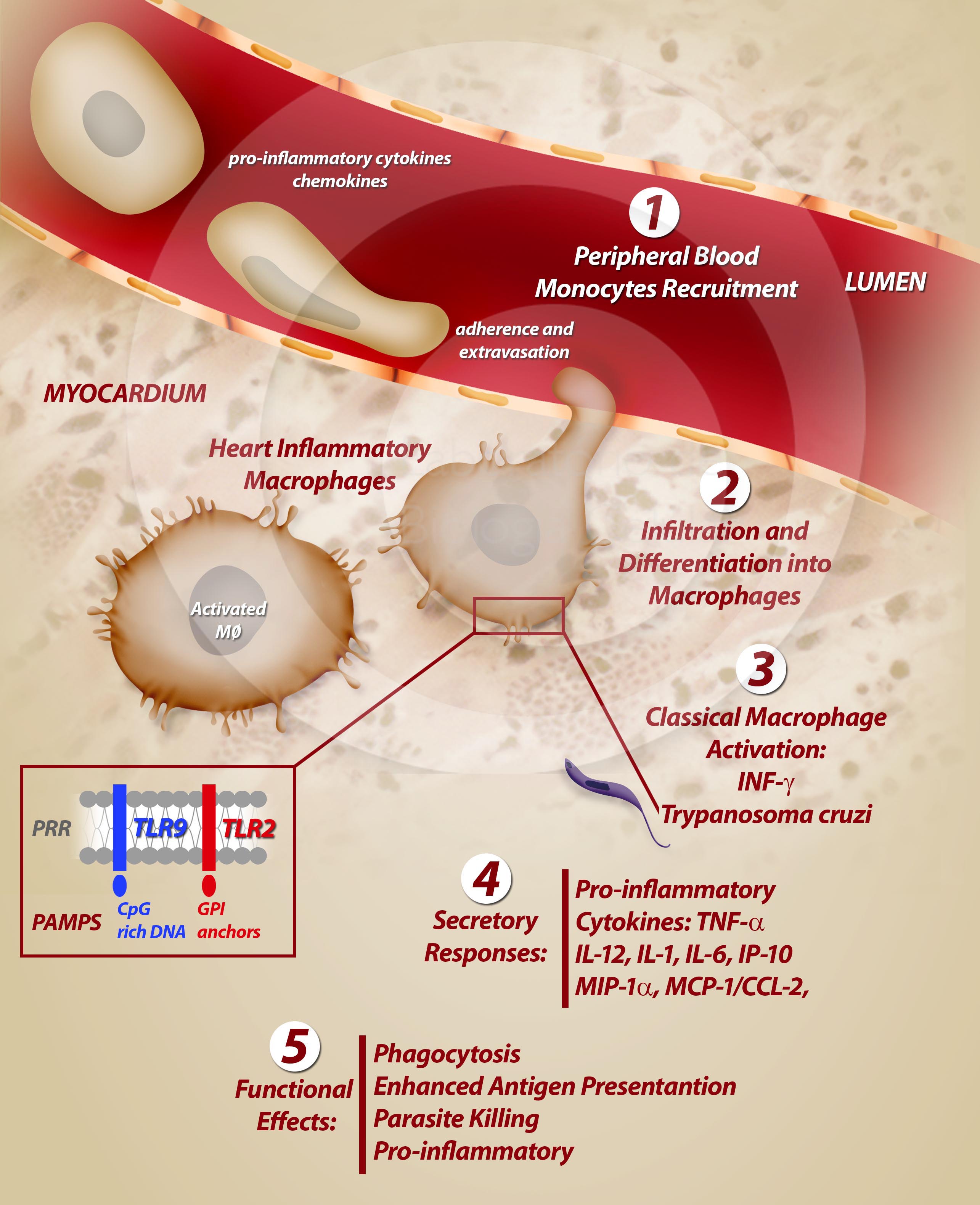
MELO, R. C. N. (2009). Acute heart inflammation: ultrastructural and functional aspects of macrophages elicited by Trypanosoma cruzi infection. J Cell Mol Med.
–